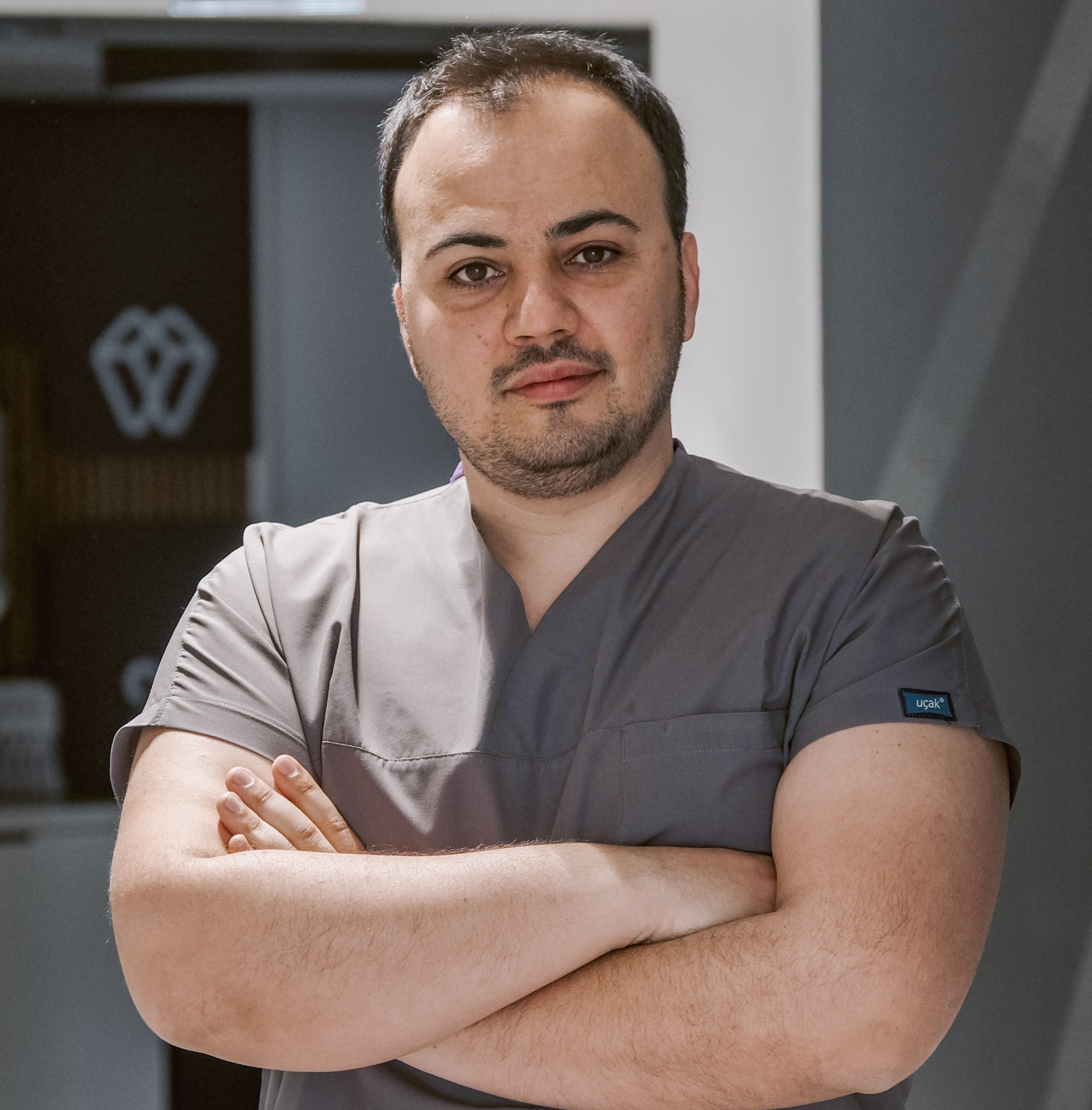
Get information about Dental Tomography treatment with the explanation of specialist dentist Mehmet Ali Koldas.
Dental Tomography

Cone Beam Computed Tomography (CBCT), Prime Dental Turkey.
Dental tomography, particularly cone beam computed tomography (CBCT), is a revolutionary imaging technique in modern dentistry and maxillofacial surgery. This technology allows for obtaining three-dimensional (3D) images of teeth, jawbones, and surrounding structures. The use of dental tomography offers significant advantages in diagnosis and treatment planning, providing comprehensive and detailed information to the practitioner. It involves obtaining a 3D representation of a 3D structure.
Dental tomography, particularly cone beam computed tomography (CBCT), is a revolutionary imaging technique in modern dentistry and maxillofacial surgery. This technology allows for obtaining three-dimensional (3D) images of teeth, jawbones, and surrounding structures. The use of dental tomography offers significant advantages in diagnosis and treatment planning, providing comprehensive and detailed information to the practitioner. It involves obtaining a 3D representation of a 3D structure.
What is Dental Tomography?
Dental tomography is an imaging technique that creates cross-sectional and three-dimensional images of the mouth and jaw structures using X-rays. CBCT is the most common form of dental tomography. The CBCT device uses a cone-shaped X-ray beam that rotates around the head, capturing images from multiple angles. These images are then processed by a computer to create a 3D model.
Uses of Dental Tomography
Dental tomography is used in diagnosing and treating various dental and medical conditions:
- It provides information such as bone density and volume necessary for the placement of dental implants. It is the best imaging method for detailed visualization in specialized implant treatments like All-on-Four and All-on-Six.
- CBCT allows for a detailed assessment of the area where implants will be placed, offering significant advantages in planning implantology treatments.
- It is highly beneficial in orthodontic treatment planning, as it evaluates tooth alignment, root angles, jaw relationships, and jaw structure.
- It shows the complex structure and issues of root canals in detail, effectively detecting lesions and fractures at the root apex.
- It is used in evaluating fractures, cracks, or other traumatic injuries, helping to determine the exact location and severity of the injury.
- It is crucial for detecting tumors, cysts, or other abnormalities within the jawbone. These structures can be visualized in 3D, aiding in treatment planning.
Advantages of Dental Tomography
- CBCT provides high-resolution, three-dimensional images of teeth, bones, and soft tissues, allowing for more accurate diagnosis and treatment planning.
- In delicate procedures like implant placement, the data obtained from CBCT enables more precise and safer surgical interventions.
- It uses a lower dose of radiation compared to traditional medical CT scans, increasing patient safety by reducing radiation exposure.
- CBCT scans are quick, usually completed within a few minutes, saving time for both the patient and the dentist.
Dental tomography has become an indispensable tool in modern dentistry and maxillofacial surgery. By providing high-resolution 3D images, it offers comprehensive information to practitioners, significantly improving the diagnosis and treatment processes. It is used in a wide range of applications, from implant planning to orthodontic evaluations, endodontic treatments to the assessment of jaw trauma. While its advantages include detailed imaging, precision, reduced radiation dose, and speed, its disadvantages such as cost and limited soft tissue imaging capabilities must also be considered. As with other conventional methods, it is not recommended to perform CBCT scans during pregnancy. Although cone beam dental tomography has a much lower radiation dose than conventional computed tomography, it has a higher radiation dose than traditional dental radiographic methods. For these reasons, it should always be requested under doctor supervision.
Mehmet Ali Koldas
He was born in 1989 in Izmir. After completing his primary, secondary and high school education in Torbalı, he went to high school in İzmir Atatürk High School.
In 2007, he started studying at Ege University, Faculty of Dentistry. In 2012, he started his specialization exam in Dentistry and started his residency in the Department of Oral and Maxillofacial Surgery at Süleyman Demirel University.


