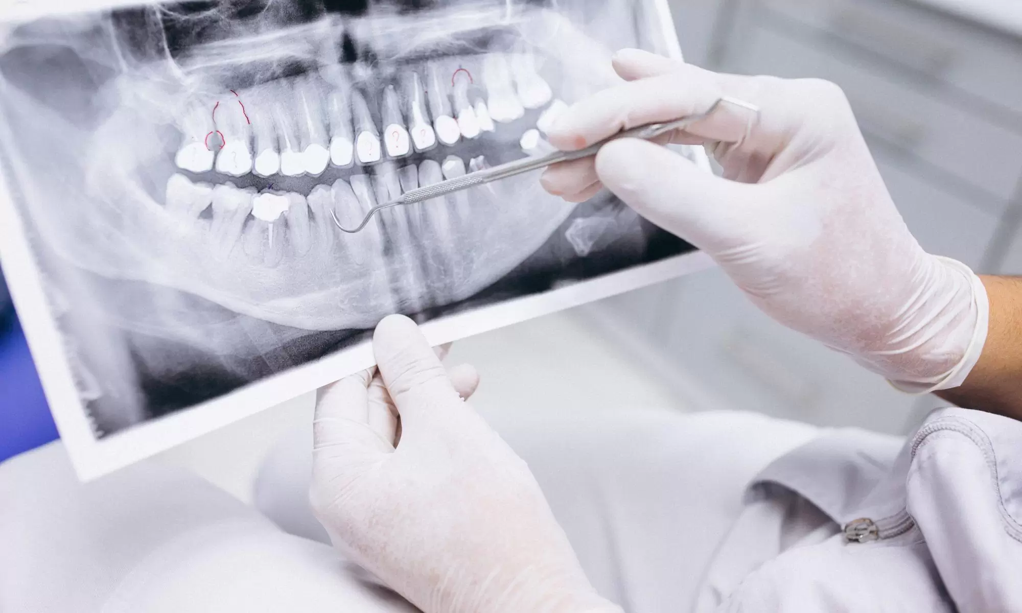
Dental X-rays are an essential tool for helping dentists diagnose issues related to teeth, jawbones, and gums. They can detect many problems, such as cavities, gum disease, root infections, and jawbone abnormalities, that are difficult to observe visually. With this information, dentists can create proper treatment plans and make necessary interventions.
What is a Dental X-ray?
A dental X-ray is a medical imaging technique used to detect and diagnose problems in the teeth and jaw area. X-rays create images of teeth and surrounding tissues.
How Does a Dental X-ray Work?
The first step is preparing the patient. The patient should remove any metal jewelry and may wear a protective apron if needed. A small film or digital sensor is placed inside the patient’s mouth to allow the X-rays to interact with the teeth.
An X-ray tube emits a beam from a specific distance next to or inside the patient’s mouth. These rays are absorbed or scattered when they pass through the teeth. X-rays are absorbed differently depending on the density of the teeth, and this contrast is used to create a black-and-white image on film or displayed directly on a screen in digital systems.
The dentist then analyzes the resulting images to detect cavities, root issues, gum diseases, or misaligned teeth.
What Can a Dental X-ray Detect?
Some common problems a dental X-ray can identify include:
- Cavities: X-rays can detect decay beneath the tooth surface, allowing early treatment.
- Root Issues: They help identify root infections, cysts, or other problems.
- Gum Disease: X-rays can reveal signs of gum disease, such as bone loss or gaps between teeth.
- Tooth Misalignment: X-rays show crowding or misalignment, aiding in orthodontic treatments.
- Gaps Between Teeth: They detect missing teeth or gaps, which helps plan treatments like implants or dentures.
Types of Dental X-rays
Dental X-rays can be taken using various techniques:
- Intraoral X-ray: Close-up images of teeth and surrounding tissues.
-
- Periapical X-ray: Provides detailed images of the tooth root and surrounding tissue.
- Bite-wing X-ray: Detects cavities between teeth and gum issues.
- Occlusal X-ray: Shows the junction between upper and lower teeth, often used in children.
- Extraoral X-ray: Used to capture images of the jaw and facial bones.
- Panoramic X-ray: Offers a general view of the mouth and jaw.
- Cephalometric X-ray: Shows the profile of the face, commonly used in orthodontics.
- CT Scan: Produces 3D images for more detailed analysis.
The appropriate technique is determined based on the patient’s needs and the dentist’s preferences.
Is Dental X-ray Safe?
Dental X-ray devices use low doses of radiation, which are safe for the body while capturing detailed images of the teeth and surrounding tissues. Patients are often given protective equipment like lead aprons to minimize exposure. Modern X-ray machines are designed to reduce radiation exposure, especially with digital systems emitting less radiation than traditional film. While the benefits usually outweigh the risks, patients should discuss their individual concerns with their dentist.
Can Dental X-rays Detect Oral Cancer?
Dental X-rays are not a direct screening tool for oral cancer but can help identify abnormalities or symptoms that may indicate cancer, such as changes in gums or the presence of tumors. A comprehensive diagnosis may require other imaging techniques and a biopsy.
How Often Should You Have a Dental X-ray?
The frequency of dental X-rays depends on the individual’s dental health, history, and risk factors. People with good dental health may only need X-rays once a year, while those with past dental issues may require them more frequently.
Is it Safe to Have a Dental X-ray Every Six Months?
Getting a dental X-ray every six months is safe for most healthy adults. Modern equipment uses very low radiation doses, and proper techniques minimize this exposure. However, individuals with certain health conditions or sensitivities to radiation may need to follow a different schedule.
Are Dental X-rays Mandatory?
Dental X-rays are often essential for diagnosing or treating specific conditions but are not mandatory. They help dentists understand a patient’s dental health and plan appropriate treatments.
Are Dental X-rays Harmful During Pregnancy?
Dental X-rays during pregnancy are usually avoided due to concerns about radiation exposure to the fetus. However, in some cases, dental X-rays may be necessary to address dental issues. If required, the dentist will take precautions to minimize radiation exposure. Alternatives to X-rays may also be considered during pregnancy. If necessary, X-rays are usually done during the second trimester when the fetus is less sensitive to radiation. Discuss any concerns with your dentist and doctor before proceeding.


